A new study from the Salk Institute identifies a brain circuit that controls voluntary breathing and emotional regulation, potentially aiding in the development of treatments for anxiety and stress disorders.
Although breathing is primarily automatic, we also possess the remarkable ability to self-soothe by slowing down our breathing. Throughout history, people have utilized slow breathing to manage emotions, with practices like mindfulness and yoga popularizing formal techniques such as box breathing. However, there has been little scientific understanding of how the brain consciously regulates our breathing and whether this has a direct impact on our anxiety and emotional state.
Neuroscience Breakthrough at Salk Institute
For the first time, neuroscientists at the Salk Institute have identified a specific brain circuit that regulates voluntary breathing. Using mice, the researchers discovered a group of neurons in the frontal cortex that connects to the brainstem, which governs essential functions like breathing. Their findings indicate that this connection between the brain’s more complex regions and the lower brainstem’s breathing center enables us to coordinate our breathing with our current behaviors and emotional state.

The study, recently published in Nature Neuroscience, describes a new set of brain cells and molecules that could be targeted with therapeutics to prevent hyperventilation and regulate anxiety, panic, or post-traumatic stress disorders.
Potential for Therapeutic Applications
“The body naturally regulates itself with deep breaths, so aligning our breathing with our emotions seems almost intuitive to us—but we didn’t really know how this worked in the brain,” says senior author Sung Han, associate professor and Pioneer Fund Developmental Chair at Salk. “By uncovering a specific brain mechanism responsible for slowing breathing, our discovery may offer a scientific explanation for the beneficial effects of practices like yoga and mindfulness on alleviating negative emotions, grounding them further in science.”
Discovering Brain Circuits for Breathing Control
Breathing patterns and emotional state are difficult to untangle—if anxiety increases or decreases, so does the breathing rate. Despite this seemingly obvious connection between emotional regulation and breathing, previous studies had only thoroughly explored subconscious breathing mechanisms in the brainstem. And while newer studies had started to describe conscious top-down mechanisms, no specific brain circuits were discovered until the Salk team took a crack at the case.
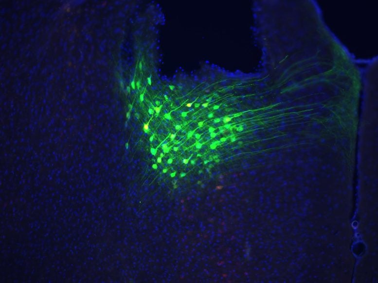
The researchers assumed the brain’s frontal cortex, which orchestrates complex thoughts and behaviors, was somehow communicating to a brainstem region called the medulla, which controls automatic breathing. To test this, they first consulted a neural connectivity database and then did experiments to trace the connections between these different brain areas.
These initial experiments revealed a potential new breathing circuit: Neurons in a frontal region called the anterior cingulate cortex were connected to an intermediate brainstem area in the pons, which was then connected to the medulla just below.
Experimental Evidence of Breathing Control
Beyond the physical connections of these brain areas, it was also important to consider the types of messages they might send each other. For example, when the medulla is active, it initiates breathing. However, messages coming down from the pons actually inhibit activity in the medulla, leading breathing rates to slow down. Han’s team hypothesized that certain emotions or behaviors could lead cortical neurons to activate the pons, which would then lower activity in the medulla, resulting in slower breath.

To test this, the researchers recorded brain activity in mice during behaviors that alter breathing, such as sniffing, swimming, and drinking, as well as during conditions that induce fear and anxiety. They also used a technique called optogenetics to turn parts of this brain circuit on or off in different emotional and behavioral contexts while measuring the animals’ breathing and behavior.
Their findings confirmed that when the connection between the cortex and the pons was activated, mice were calmer and breathed more slowly, but when mice were in anxiety-inducing situations, this communication decreased, and breathing rates went up. Furthermore, when the researchers artificially activated this cortex-pons-medulla circuit, the animals’ breath slowed, and they showed fewer signs of anxiety. On the other hand, if researchers shut this circuit off, breathing rates went up, and the mice became more anxious.
Altogether, this anterior cingulate cortex-pons-medulla circuit supported the voluntary coordination of breathing rates with behavioral and emotional states.
Implications and Future Research Directions
“Our findings got me thinking: Could we develop drugs to activate these neurons and manually slow our breathing or prevent hyperventilation in panic disorder?” says the first author of the study, Jinho Jhang, a senior research associate in Han’s lab. “My sister, three years younger than me, has suffered from panic disorder for many years. She continues to inspire my research questions and my dedication to answering them.”
The researchers will continue analyzing the circuit to determine whether drugs could activate it to slow breathing on command. Additionally, the team is working to find the circuit’s converse—a fast-breathing circuit, which they believe is likely also tied to emotion. They are hopeful their findings will result in long-term solutions for people with anxiety, stress, and panic disorders, who inspire their discovery and dedication.
“I want to use these findings to design a yoga pill,” says Han. “It may sound silly, and the translation of our work into a marketable drug will take years, but we now have a potentially targetable brain circuit for creating therapeutics that could instantly slow breathing and initiate a peaceful, meditative state.”
Reference: “A top-down slow breathing circuit that alleviates negative affect in mice” by Jinho Jhang, Seahyung Park, Shijia Liu, David D. O’Keefe and Sung Han, 19 November 2024, Nature Neuroscience.
DOI: 10.1038/s41593-024-01799-w
The work was supported by the Kavli Institute for Brain and Mind (IRGS 2020-1710).
This post was originally published on here


