This post was originally published on here
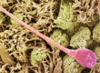
Tiny plastic fragments known as microplastics and nanoplastics have spread across the planet. They have been found in deep ocean waters, farmland soils, wildlife, and even inside the human body. Despite their widespread presence, researchers still do not fully understand what happens after these particles enter living organisms. A new study outlines a fluorescence-based technique that could allow scientists to monitor microplastics in real time as they move through the body, change chemically, and eventually break down.
Plastic production worldwide now surpasses 460 million tons per year. Each year, millions of tons of microscopic plastic particles are released into the environment. Scientists have identified these particles in marine animals, birds, and human tissues including blood, liver, and even brain samples. Laboratory experiments suggest exposure may be linked to inflammation, organ damage, and developmental problems. Even so, a critical knowledge gap remains about how these particles behave once inside living systems.
“Most current methods give us only a snapshot in time,” said corresponding author Wenhong Fan. “We can measure how many particles are present in a tissue, but we cannot directly observe how they travel, accumulate, transform, or break down inside living organisms.”
Limits of Current Microplastic Detection Methods
Common detection tools such as infrared spectroscopy and mass spectrometry require scientists to destroy tissue samples in order to analyze them. This approach prevents researchers from watching how particles behave over time. Fluorescence imaging offers a possible solution, but current labeling techniques often face problems such as fading signals, leaking dyes, or reduced brightness in complex biological environments.
A New Fluorescent Strategy for Real-Time Tracking
To address these limitations, the team designed what they call a fluorescent monomer controlled synthesis strategy. Rather than coating plastic particles with fluorescent dye, they incorporated light-emitting components directly into the plastic’s molecular structure. The method uses aggregation induced emission materials, which glow more intensely when clustered together. This design helps maintain a stable signal and reduces the loss of brightness during imaging.
With this technique, researchers can fine-tune particle brightness, color of emitted light, size, and shape. Because the fluorescent material is evenly distributed throughout each particle, both whole plastics and the smaller fragments created as they degrade remain visible. That capability opens the door to tracking the full life cycle of microplastics, from ingestion and internal transport to transformation and final breakdown.
Understanding Health and Environmental Risks
The strategy is still being tested experimentally, but it is based on established principles from polymer chemistry and biocompatible fluorescence imaging. The researchers say the approach could become an important tool for studying how microplastics interact with cells, tissues, and organs.
“Clarifying the transport and transformation processes of microplastics inside organisms is essential for assessing their true ecological and health risks,” Fan said. “Dynamic tracking will help us move beyond simple exposure measurements toward a deeper understanding of toxicity mechanisms.”
As worries about plastic pollution intensify, tools that reveal how microplastics behave inside living systems may play a key role in improving risk assessments and guiding future environmental regulations.